anti-AIF1 / Iba 1 antibody
| 产品描述 | Rabbit Polyclonal antibody recognizes AIF1 / Iba 1 |
|---|---|
| 反应物种 | Hu, Ms, Rat |
| 应用 | ICC/IF, IHC-Fr, WB |
| 宿主 | Rabbit |
| 克隆 | Polyclonal |
| 同位型 | IgG |
| 靶点名称 | AIF1 / Iba 1 |
| 抗原物种 | Human |
| 抗原 | KLH-conjugated synthetic peptide around the C-terminal region of Human AIF1 / Iba1. |
| 偶联标记 | Un-conjugated |
| 別名 | Ionized calcium-binding adapter molecule 1; Allograft inflammatory factor 1; IBA1; IRT-1; IRT1; AIF-1; Protein G1 |
| 应用建议 |
| ||||||||
|---|---|---|---|---|---|---|---|---|---|
| 应用说明 | * The dilutions indicate recommended starting dilutions and the optimal dilutions or concentrations should be determined by the scientist. | ||||||||
| 实际分子量 | 17 kDa |
| 形式 | Liquid |
|---|---|
| 纯化 | Unpurified |
| 缓冲液 | Serum and 5mM Sodium azide. |
| 抗菌剂 | 5mM Sodium azide |
| 存放说明 | For continuous use, store undiluted antibody at 2-8°C for up to a week. For long-term storage, aliquot and store at -20°C or below. Storage in frost free freezers is not recommended. Avoid repeated freeze/thaw cycles. Suggest spin the vial prior to opening. The antibody solution should be gently mixed before use. |
| 注意事项 | For laboratory research only, not for drug, diagnostic or other use. |
| 数据库连接 | |
|---|---|
| 基因名称 | AIF1 |
| 全名 | allograft inflammatory factor 1 |
| 背景介绍 | AIF1 / Iba 1 is a protein that binds actin and calcium. This gene is induced by cytokines and interferon and may promote macrophage activation and growth of vascular smooth muscle cells and T-lymphocytes. Polymorphisms in this gene may be associated with systemic sclerosis. Alternative splicing results in multiple transcript variants, but the full-length and coding nature of some of these variants is not certain. [provided by RefSeq, Jan 2016] |
| 生物功能 | AIF1 / Iba 1 is an Actin-binding protein. It enhances membrane ruffling and RAC activation. Enhances the actin-bundling activity of LCP1. Binds calcium. Plays a role in RAC signaling and in phagocytosis. May play a role in macrophage activation and function. Promotes the proliferation of vascular smooth muscle cells and of T-lymphocytes. Enhances lymphocyte migration. Plays a role in vascular inflammation. [UniProt] |
| 产品亮点 | Related Antibody Duos and Panels: ARG30324 Neuroinflammation Antibody Panel Related products: AIF1 antibodies; AIF1 Duos / Panels; Anti-Rabbit IgG secondary antibodies; |
| 研究领域 | Cell Biology and Cellular Response antibody; Immune System antibody; Metabolism antibody; Neuroscience antibody; Activated Macrophage/Microglia Study antibody; Neuroinflammation Study antibody; Macroglial Marker antibody |
| 预测分子量 | 17 kDa |
| 翻译后修饰 | Phosphorylated on serine residues. [UniProt] |
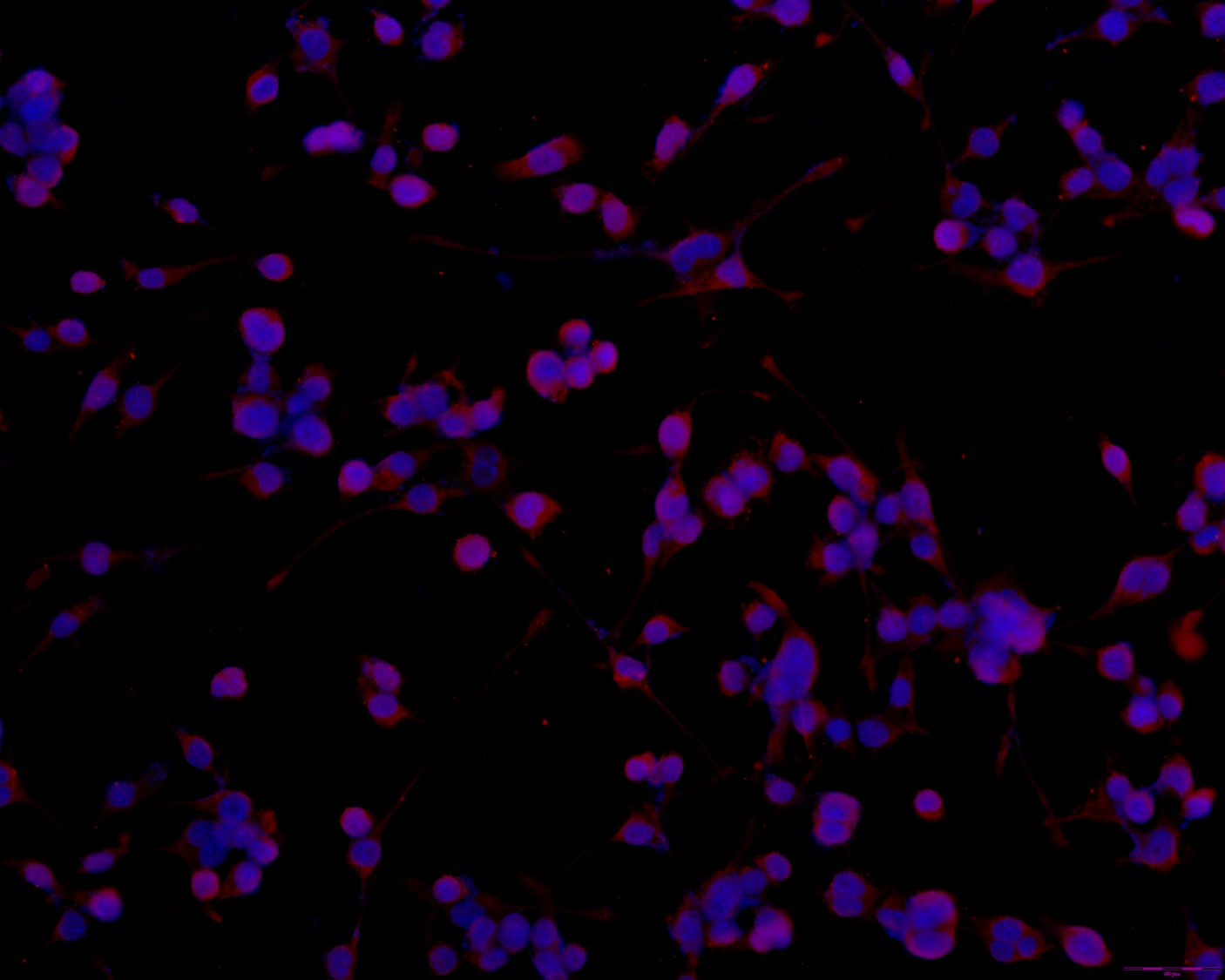
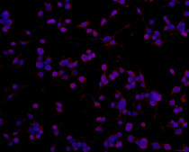
ARG11062 anti-AIF1 / Iba 1 antibody ICC/IF image (Customer's Feedback)
Immunofluorescence: BV-2 stained with ARG11062 anti-AIF1 / Iba 1 antibody at 1:100 dilution.
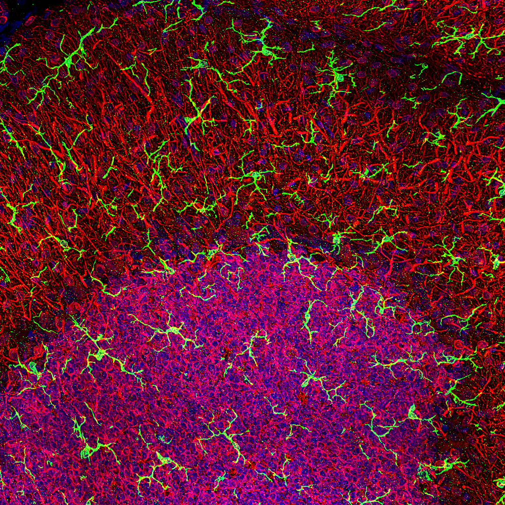
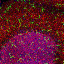
ARG11062 anti-AIF1 / Iba 1 antibody IHC-Fr image
Immunohistochemistry: High magnification stacked confocal image of Rat cerebellar molecular layer at top and granular layer below, stained with ARG11062 anti-AIF1 / Iba 1 antibody (green) at 1:1000 dilution. Nuclear DNA is shown with DAPI stain in blue.
Microglia are very small cells with fine processes spreading in three dimensions and so are best visualized in a confocal Z stack. Red shows the processes of Purkinje cells and the perikarya of granule cells revealed by an antibody to MAP2, at 1:5000 dilution.
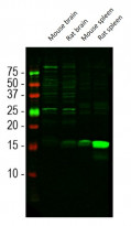
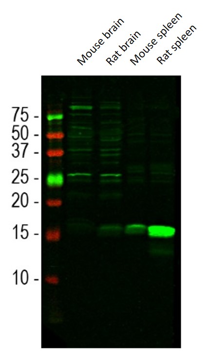
ARG11062 anti-AIF1 / Iba 1 antibody WB image
Western blot: Mouse brain, Rat brain, Mouse spleen, and Rat spleen lysates stained with ARG11062 anti-AIF1 / Iba 1 antibody at 1:1000 dilution.
Iba1 is a relatively minor protein of brain and is much more abundant in spleen, so the 15 kDa band is less obvious in CNS lysates. The other bands seen in the CNS lysates are of unknown origin but do not appear to compromise the migroglial specific staining seen with this antibody.
 New Products


