anti-Aldolase C antibody [4A9]
| 产品描述 | Mouse Monoclonal antibody [4A9] recognizes Aldolase C |
|---|---|
| 反应物种 | Hu, Ms, Rat, Cow, Hrs, Pig |
| 预测物种 | Chk |
| 应用 | ICC/IF, IHC-Fr, WB |
| 宿主 | Mouse |
| 克隆 | Monoclonal |
| 克隆号 | 4A9 |
| 同位型 | IgG1 |
| 靶点名称 | Aldolase C |
| 抗原 | N-terminal sequence MPHSYPALSAEQKKELSDIA |
| 偶联标记 | Un-conjugated |
| 別名 | EC 4.1.2.13; Brain-type aldolase; Fructose-bisphosphate aldolase C; ALDC |
| 应用建议 |
| ||||||||
|---|---|---|---|---|---|---|---|---|---|
| 应用说明 | * The dilutions indicate recommended starting dilutions and the optimal dilutions or concentrations should be determined by the scientist. |
| 形式 | Liquid |
|---|---|
| 纯化 | Affinity purification. |
| 缓冲液 | PBS and 50% Glycerol. |
| 稳定剂 | 50% Glycerol |
| 浓度 | 1 mg/ml |
| 存放说明 | For continuous use, store undiluted antibody at 2-8°C for up to a week. For long-term storage, aliquot and store at -20°C. Storage in frost free freezers is not recommended. Avoid repeated freeze/thaw cycles. Suggest spin the vial prior to opening. The antibody solution should be gently mixed before use. |
| 注意事项 | For laboratory research only, not for drug, diagnostic or other use. |
| 数据库连接 | |
|---|---|
| 基因名称 | ALDOC |
| 全名 | aldolase C, fructose-bisphosphate |
| 背景介绍 | This gene encodes a member of the class I fructose-biphosphate aldolase gene family. Expressed specifically in the hippocampus and Purkinje cells of the brain, the encoded protein is a glycolytic enzyme that catalyzes the reversible aldol cleavage of fructose-1,6-biphosphate and fructose 1-phosphate to dihydroxyacetone phosphate and either glyceraldehyde-3-phosphate or glyceraldehyde, respectively. [provided by RefSeq, Jul 2008] |
| 预测分子量 | 39 kDa |
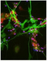
ARG10695 anti-Aldolase C antibody [4A9] ICC/IF image
Immunocytochemistry: Rat mixed neuron / glial cultures stained with ARG10695 anti-Aldolase C antibody [4A9] (green) and co-stained with our rabbit antibody to FOX3 / NeuN (red). ARG10695 anti-Aldolase C antibody [4A9] antibody reveals strong cytoplasmic staining in astrocytes, while Rabbit FOX3 / NeuN antibody shows nuclear and distal cytoplasmic staining in neuron cells and is complete absence of astrocytes. Blue is a DNA stain.
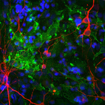
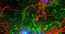
ARG10695 anti-Aldolase C antibody [4A9] ICC/IF image
Immunofluorescence: Cortical neuron-glial cells from E20 Rat stained with ARG10695 anti-Aldolase C antibody [4A9] (green) at 1:1000 dilution and costained with ARG52328 anti-MAP2 antibody (red) at 1:10000 dilution. Hoechst (blue) for nuclear staining.
The Aldolase C antibody labels cytosolic protein expressed in the glial cells, while the MAP2 antibody stains dendrites and perikarya of mature neurons.
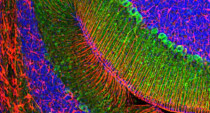
ARG10695 anti-Aldolase C antibody [4A9] IHC-Fr image
Immunohistochemistry: Frozen section of Rat cerebellum stained with ARG10695 anti-Aldolase C antibody [4A9] (green) at 1:1000 dilution and costained with ARG52312 anti-GFAP antibody (red) at 1:5000 dilution. Hoechst (blue) for nuclear staining.
Aldolase C antibody selectively labels the perikarya and dendrites of Purkinje cells, while GFAP antibody stains processes of Bergman glia and astrocytic cells.
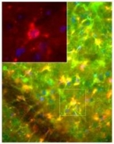
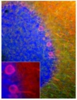
ARG10695 anti-Aldolase C antibody [4A9] IHC-Fr image
Immunohistochemistry: Frozen sections of Mouse brain (fixed by transcardial perfusion with 4% paraformaldehyde) stained with ARG10695 anti-Aldolase C antibody [4A9] (red) and co-stained with chicken anti-Vimentin antibody (green). In the striatum, ARG10695 anti-Aldolase C antibody [4A9] positive astrocytes are highly co-stained with Vimentin antibody, which results in yellow to gold colors. Insets show a higher magnification picture. Nuclei are labeled with DAPI (blue).
ARG10695 anti-Aldolase C antibody [4A9] WB image
Western blot: Crude cow cerebellum homogenate stained with ARG10695 anti-Aldolase C antibody [4A9]. Other studies show that this antibody does not recognize the very closely related molecules aldolase A or B.
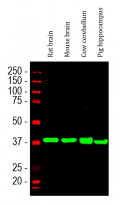
ARG10695 anti-Aldolase C antibody [4A9] WB image
Western blot: Rat brain, Mouse brain, Cow cerebellum and Pig hippocampus lysates stained with ARG10695 anti-Aldolase C antibody [4A9] (green) at 1:2000 dilution.
ARG10695 anti-Aldolase C antibody [4A9] IHC-Fr image
Immunohistochemistry: Frozen section of E20 Rat cortical tissue stained with ARG10695 anti-Aldolase C antibody [4A9] (green) at 1:1000 dilution and costained with ARG52328 anti-MAP2 antibody (red) at 1:10000 dilution.
The aldolase C antibody labels cytosolic protein expressed in glial cells, while MAP2 antibody stains dendrites and perikarya of mature neurons.
ARG10695 anti-Aldolase C antibody [4A9] IHC-Fr image
Immunohistochemistry: Frozen sections of Mouse brain (fixed by transcardial perfusion with 4% paraformaldehyde) stained with ARG10695 anti-Aldolase C antibody [4A9] (red) and co-stained with chicken anti-Vimentin antibody (green). In the cerebellum, ARG10695 anti-Aldolase C antibody [4A9] positive Purkinje cells do not express vimentin, which results in red color. Insets show a higher magnification picture. Nuclei are labeled with DAPI (blue).
ARG10695 anti-Aldolase C antibody [4A9] WB image
Western blot: Recombinant Human Aldolase A, B and C with three monoclonal antibodies as indicated showing binding to all three gene products. Lane labelled S show molecular weight standards, while lanes A, B and C contain recombinant full length His-tagged Human Aldolase A, B and C respectively. The epitope for ARG10695 anti-Aldolase C antibody [4A9] is within the N-terminal peptide of the molecule which is less conserved between the three Aldolases. Clone 1A1 binds to the C-terminal peptide and also is specific for Aldolase C, while clone E9 binds to the less conserved core of the molecule and recognizes all three Aldolase C gene products.
 New Products


![anti-Aldolase C antibody [4A9]](/upload/image/products/ARG10695_WB_2_210_205.jpg)
![anti-Aldolase C antibody [4A9]](/upload/image/products/ARG10695_ICC_1.jpg)
![anti-Aldolase C antibody [4A9]](/upload/image/products/ARG10695_IF_190729.jpg)
![anti-Aldolase C antibody [4A9]](/upload/image/products/ARG10695_IHC-Fr_190729-1.jpg)
![anti-Aldolase C antibody [4A9]](/upload/image/products/ARG10695_IHC_1.jpg)
![anti-Aldolase C antibody [4A9]](/upload/image/products/ARG10695_WB_1.jpg)
![anti-Aldolase C antibody [4A9]](/upload/image/products/ARG10695_WB_190729.jpg)
![anti-Aldolase C antibody [4A9]](/upload/image/products/ARG10695_IHC-Fr_190729-2.jpg)
![anti-Aldolase C antibody [4A9]](/upload/image/products/ARG10695_IHC_2.jpg)
![anti-Aldolase C antibody [4A9]](/upload/image/products/ARG10695_WB_2.jpg)
