anti-APE1 antibody
CAT.NO. : ARG63219
US$ Please choose
US$ Please choose
Size:
Trail, Bulk size or Custom requests Please contact us
概述
| 产品描述 | Goat Polyclonal antibody recognizes APE1 |
|---|---|
| 反应物种 | Hu |
| 预测物种 | Cow, Dog, Pig |
| 应用 | IHC-P, WB |
| 特异性 | Reported variants represent identical protein (NP_001632.2; NP_542379.1; NP_542380.1). |
| 宿主 | Goat |
| 克隆 | Polyclonal |
| 同位型 | IgG |
| 靶点名称 | APE1 |
| 抗原物种 | Human |
| 抗原 | PKRGKKGAVAEDGD-C |
| 偶联标记 | Un-conjugated |
| 別名 | APE1; APX; AP endonuclease 1; HAP1; EC 4.2.99.18; Apurinic-apyrimidinic endonuclease 1; APEX; apurinic or apyrimidinic site; EC 3.1.-.-; REF1; Redox factor-1; APEX nuclease; REF-1; APE-1; DNA-; APEN; APE |
应用说明
| 应用建议 |
| ||||||
|---|---|---|---|---|---|---|---|
| 应用说明 | IHC-P: Antigen Retrieval: Steam tissue section in Tris/EDTA buffer (pH 9.0). WB: Recommend incubate at RT for 1h. * The dilutions indicate recommended starting dilutions and the optimal dilutions or concentrations should be determined by the scientist. |
属性
| 形式 | Liquid |
|---|---|
| 纯化 | Purified from goat serum by ammonium sulphate precipitation followed by antigen affinity chromatography using the immunizing peptide. |
| 缓冲液 | Tris saline (pH 7.3), 0.02% Sodium azide and 0.5% BSA |
| 抗菌剂 | 0.02% Sodium azide |
| 稳定剂 | 0.5% BSA |
| 浓度 | 0.5 mg/ml |
| 存放说明 | For continuous use, store undiluted antibody at 2-8°C for up to a week. For long-term storage, aliquot and store at -20°C or below. Storage in frost free freezers is not recommended. Avoid repeated freeze/thaw cycles. Suggest spin the vial prior to opening. The antibody solution should be gently mixed before use. |
| 注意事项 | For laboratory research only, not for drug, diagnostic or other use. |
生物信息
| 数据库连接 | Swiss-port # P27695 Human DNA-(apurinic or apyrimidinic site) lyase |
|---|---|
| 基因名称 | APEX1 |
| 全名 | APEX nuclease (multifunctional DNA repair enzyme) 1 |
| 背景介绍 | Apurinic/apyrimidinic (AP) sites occur frequently in DNA molecules by spontaneous hydrolysis, by DNA damaging agents or by DNA glycosylases that remove specific abnormal bases. AP sites are pre-mutagenic lesions that can prevent normal DNA replication so the cell contains systems to identify and repair such sites. Class II AP endonucleases cleave the phosphodiester backbone 5' to the AP site. This gene encodes the major AP endonuclease in human cells. Splice variants have been found for this gene; all encode the same protein. [provided by RefSeq, Jul 2008] |
| 研究领域 | Cell Biology and Cellular Response antibody; Gene Regulation antibody |
| 预测分子量 | 36 kDa |
| 翻译后修饰 | Phosphorylated. Phosphorylation by kinase PKC or casein kinase CK2 results in enhanced redox activity that stimulates binding of the FOS/JUN AP-1 complex to its cognate binding site. AP-endodeoxyribonuclease activity is not affected by CK2-mediated phosphorylation. Phosphorylation of Thr-233 by CDK5 reduces AP-endodeoxyribonuclease activity resulting in accumulation of DNA damage and contributing to neuronal death. Acetylated on Lys-6 and Lys-7. Acetylation is increased by the transcriptional coactivator EP300 acetyltransferase, genotoxic agents like H(2)O(2) and methyl methanesulfonate (MMS). Acetylation increases its binding affinity to the negative calcium response element (nCaRE) DNA promoter. The acetylated form induces a stronger binding of YBX1 to the Y-box sequence in the MDR1 promoter than the unacetylated form. Deacetylated on lysines. Lys-6 and Lys-7 are deacetylated by SIRT1. Cleaved at Lys-31 by granzyme A to create the mitochondrial form; leading in reduction of binding to DNA, AP endodeoxynuclease activity, redox activation of transcription factors and to enhanced cell death. Cleaved by granzyme K; leading to intracellular ROS accumulation and enhanced cell death after oxidative stress. Cys-65 and Cys-93 are nitrosylated in response to nitric oxide (NO) and lead to the exposure of the nuclear export signal (NES). Ubiquitinated by MDM2; leading to translocation to the cytoplasm and proteasomal degradation. |
检测图片 (2)
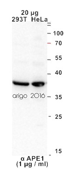
ARG63219 anti-APE1 antibody WB image
Western blot: 20 μg of 1) 293T and 2) HeLa cell lysates stained with ARG63219 anti-APE1 antibody at 1 μg/ml dilution.
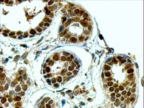
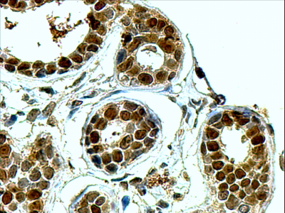
ARG63219 anti-APE1 antibody IHC image
Immunohistochemistry: paraffin-embedded Human Breast. (Steamed antigen retrieval with Tris/EDTA buffer pH 9) stained with ARG63219 anti-APE1 antibody at 4 µg/ml dilution, followed by HRP-staining.
 New Products


