anti-Ataxin 1 antibody [S65-37]
| 产品描述 | Mouse Monoclonal antibody [S65-37] recognizes Ataxin 1 |
|---|---|
| 反应物种 | Hu, Ms, Rat |
| 应用 | ICC/IF, WB |
| 特异性 | Detects ~85kDa. No cross-reactivity against phosphor S751-Ataxin-1. Minimal cross-reactivity against S751A mutant of Ataxin-1 by ELISA and immunofluorescence and negative by immunoblot. |
| 宿主 | Mouse |
| 克隆 | Monoclonal |
| 克隆号 | S65-37 |
| 同位型 | IgG1 |
| 靶点名称 | Ataxin 1 |
| 抗原物种 | Mouse |
| 抗原 | Synthetic peptide around aa. 746-761 (RKRRWSAPETRKLEKS) of Mouse ataxin 1. Rat: 93% identity (15/16 amino acids identical). Human: 87% identity (14/16 amino acids identical). |
| 偶联标记 | Un-conjugated |
| 別名 | SCA1; D6S504E; ATX1; Ataxin-1; Spinocerebellar ataxia type 1 protein |
| 应用建议 |
| ||||||
|---|---|---|---|---|---|---|---|
| 应用说明 | * The dilutions indicate recommended starting dilutions and the optimal dilutions or concentrations should be determined by the scientist. |
| 形式 | Liquid |
|---|---|
| 纯化 | Purification with Protein G. |
| 缓冲液 | PBS (pH 7.4), 0.1% Sodium azide and 50% Glycerol |
| 抗菌剂 | 0.1% Sodium azide |
| 稳定剂 | 50% Glycerol |
| 浓度 | 1 mg/ml |
| 存放说明 | For continuous use, store undiluted antibody at 2-8°C for up to a week. For long-term storage, aliquot and store at -20°C. Storage in frost free freezers is not recommended. Avoid repeated freeze/thaw cycles. Suggest spin the vial prior to opening. The antibody solution should be gently mixed before use. |
| 注意事项 | For laboratory research only, not for drug, diagnostic or other use. |
| 数据库连接 | |
|---|---|
| 基因名称 | Atxn1 |
| 全名 | ataxin 1 |
| 背景介绍 | The autosomal dominant cerebellar ataxias (ADCA) are a heterogeneous group of neurodegenerative disorders characterized by progressive degeneration of the cerebellum, brain stem and spinal cord. Clinically, ADCA has been divided into three groups: ADCA types I-III. ADCAI is genetically heterogeneous, with five genetic loci, designated spinocerebellar ataxia (SCA) 1, 2, 3, 4 and 6, being assigned to five different chromosomes. ADCAII, which always presents with retinal degeneration (SCA7), and ADCAIII often referred to as the `pure' cerebellar syndrome (SCA5), are most likely homogeneous disorders. Several SCA genes have been cloned and shown to contain CAG repeats in their coding regions. ADCA is caused by the expansion of the CAG repeats, producing an elongated polyglutamine tract in the corresponding protein. The expanded repeats are variable in size and unstable, usually increasing in size when transmitted to successive generations. The function of the ataxins is not known. This locus has been mapped to chromosome 6, and it has been determined that the diseased allele contains 41-81 CAG repeats, compared to 6-39 in the normal allele, and is associated with spinocerebellar ataxia type 1 (SCA1). At least two transcript variants encoding the same protein have been found for this gene. [provided by RefSeq, Jan 2010] |
| 生物功能 | Chromatin-binding factor that repress Notch signaling in the absence of Notch intracellular domain by acting as a CBF1 corepressor. Binds to the HEY promoter and might assist, along with NCOR2, RBPJ-mediated repression. Binds RNA in vitro. May be involved in RNA metabolism. [UniProt] |
| 细胞定位 | Cytoplasm, Nucleus |
| 预测分子量 | 87 kDa |
| 翻译后修饰 | Ubiquitinated by UBE3A, leading to its degradation by the proteasome. The presence of expanded poly-Gln repeats in spinocerebellar ataxia 1 (SCA1) patients impairs ubiquitination and degradation, leading to accumulation of ATXN1 in neurons and subsequent toxicity. Phosphorylation at Ser-775 increases the pathogenicity of proteins with an expanded polyglutamine tract. Sumoylation is dependent on nuclear localization and phosphorylation at Ser-775. It is reduced in the presence of an expanded polyglutamine tract. |
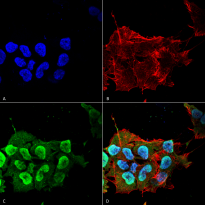
ARG22254 anti-Ataxin 1 antibody [S65-37] ICC/IF image
Immunofluorescence: Human Neuroblastoma cell line SK-N-BE. Fixation: 4% Formaldehyde for 15 min at RT. Primary Antibody: ARG22254 anti-Ataxin 1 antibody [S65-37] at 1:100 for 60 min at RT. Secondary Antibody: Goat anti-Mouse ATTO 488 at 1:100 for 60 min at RT. Counterstain: Phalloidin Texas Red F-Actin stain; DAPI (blue) nuclear stain. Magnification: 60X. (A) DAPI (blue) nuclear stain (B) Phalloidin Texas Red F-Actin stain (C) ARG22254 anti-Ataxin 1 antibody [S65-37] (D) Composite.
ARG22254 anti-Ataxin 1 antibody [S65-37] WB image
Western blot: 15 µg of Monkey COS-1 cells transfected with Ataxin-1. Block: 2% BSA and 2% Skim Milk in 1X TBST. Primary Antibody: ARG22254 anti-Ataxin 1 antibody [S65-37] at 1:200 for 16 hours at 4°C. Secondary Antibody: Goat anti-Mouse IgG: HRP at 1:1000 for 1 hour RT. Color Development: ECL solution for 6 min in RT.
 New Products


![anti-Ataxin 1 antibody [S65-37]](/upload/image/products/ARG22254_WB-Monkey-COS-1_210_205.jpg)
![anti-Ataxin 1 antibody [S65-37]](/upload/image/products/ARG22254_ICC-IF-Human-Neuroblastoma.jpg)
![anti-Ataxin 1 antibody [S65-37]](/upload/image/products/ARG22254_WB-Monkey-COS-1.jpg)
