anti-IL1 beta antibody [SQab1748]
| 产品描述 | Mouse Monoclonal antibody [SQab1748] recognizes IL1 beta |
|---|---|
| 反应物种 | Hu, Ms |
| 应用 | ELISA, FACS, ICC/IF, IHC-P, WB |
| 特异性 | Not cross-react with recombinant human IL-1 alpha in ELISA (5 µg/ml protein) and WB (100 ng protein). |
| 宿主 | Mouse |
| 克隆 | Monoclonal |
| 克隆号 | SQab1748 |
| 同位型 | IgG2a |
| 靶点名称 | IL1 beta |
| 抗原物种 | Human |
| 抗原 | Recombinant Human IL1 beta protein. |
| 偶联标记 | Un-conjugated |
| 別名 | Interleukin-1 beta; IL1-BETA; IL-1; IL-1 beta; Catabolin; IL1F2 |
| 应用建议 |
| ||||||||||||
|---|---|---|---|---|---|---|---|---|---|---|---|---|---|
| 应用说明 | * The dilutions indicate recommended starting dilutions and the optimal dilutions or concentrations should be determined by the scientist. |
| 形式 | Liquid |
|---|---|
| 缓冲液 | PBS (pH 7.4) and 0.01% Thimerosal. |
| 抗菌剂 | 0.01% Thimerosal |
| 浓度 | 1 mg/ml |
| 存放说明 | For continuous use, store undiluted antibody at 2-8°C for up to a week. For long-term storage, aliquot and store at -20°C or below. Storage in frost free freezers is not recommended. Avoid repeated freeze/thaw cycles. Suggest spin the vial prior to opening. The antibody solution should be gently mixed before use. |
| 注意事项 | For laboratory research only, not for drug, diagnostic or other use. |
| 数据库连接 | |
|---|---|
| 基因名称 | IL1B |
| 全名 | interleukin 1, beta |
| 背景介绍 | IL1 beta protein is a member of the interleukin 1 cytokine family. This cytokine is produced by activated macrophages as a proprotein, which is proteolytically processed to its active form by caspase 1 (CASP1/ICE). This cytokine is an important mediator of the inflammatory response, and is involved in a variety of cellular activities, including cell proliferation, differentiation, and apoptosis. The induction of cyclooxygenase-2 (PTGS2/COX2) by this cytokine in the central nervous system (CNS) is found to contribute to inflammatory pain hypersensitivity. This gene and eight other interleukin 1 family genes form a cytokine gene cluster on chromosome 2. [provided by RefSeq, Jul 2008] |
| 生物功能 | IL1 beta is a potent proinflammatory cytokine. Initially discovered as the major endogenous pyrogen, induces prostaglandin synthesis, neutrophil influx and activation, T-cell activation and cytokine production, B-cell activation and antibody production, and fibroblast proliferation and collagen production. Promotes Th17 differentiation of T-cells. Synergizes with IL12/interleukin-12 to induce IFNG synthesis from T-helper 1 (Th1) cells (PubMed:10653850). [UniProt] |
| 产品亮点 | Related Antibody Duos and Panels: ARG30330 Pyroptosis Antibody Panel Related products: IL1 beta antibodies; IL1 beta ELISA Kits; IL1 beta Duos / Panels; IL1 beta recombinant proteins; Anti-Mouse IgG secondary antibodies; Related news: HMGB1 in inflammation Inflammatory Cytokines Exploring Antiviral Immune Response RIP1 activation and pathogenesis of NASH |
| 研究领域 | Pyroptosis Study antibody |
| 预测分子量 | 31 kDa |
| 翻译后修饰 | Activation of the IL1B precursor involves a CASP1-catalyzed proteolytic cleavage. Processing and secretion are temporarily associated. [UniProt] |
ARG66285 anti-IL1 beta antibody [SQab1748] WB image
Western blot: 20 μg of A431 and HeLa cell lysates stained with ARG66285 anti-IL1 beta antibody [SQab1748] at 1:4000 dilution.
ARG66285 anti-IL1 beta antibody [SQab1748] ICC/IF image
Immunofluorescence: MCF7 cells stained with ARG66285 anti-IL1 beta antibody [SQab1748] at 1:200 dilution.
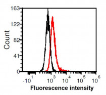
ARG66285 anti-IL1 beta antibody [SQab1748] FACS image
Flow Cytometry: HeLa cells stained with ARG66285 anti-IL1 beta antibody [SQab1748] at 1 µg/ml (red) and without antibody control (black).
ARG66285 anti-IL1 beta antibody [SQab1748] ICC/IF image
Immunofluorescence: HeLa cells were fixed in 4% PFA, permeabilized with PBS containing 0.1% Triton X-100. Cells were stained with ARG66285 anti-IL1 beta antibody [SQab1748] (green) at 1:200 dilution, and cell nuclei were stained with Hoechst 33342 (blue).
ARG66285 anti-IL1 beta antibody [SQab1748] WB image
Western blot: 50 µg of Mouse liver lysate stained with ARG66285 anti-IL1 beta antibody [SQab1748] at 1:5000 dilution.
ARG66285 anti-IL1 beta antibody [SQab1748] WB image
Western blot: 50 µg of Rat liver lysate stained with ARG66285 anti-IL1 beta antibody [SQab1748] at 1:5000 dilution.
 New Products


![anti-IL1 beta antibody [SQab1748]](/upload/image/products/ARG66285_WB_RatLiver_210_205.jpg)
![anti-IL1 beta antibody [SQab1748]](/upload/image/products/ARG66285_WB_SheetRev_A431_HeLa_191206.jpg)
![anti-IL1 beta antibody [SQab1748]](/upload/image/products/ARG66285_IF_MCF7.jpg)
![anti-IL1 beta antibody [SQab1748]](/upload/image/products/ARG66285_FACS_HeLa.jpg)
![anti-IL1 beta antibody [SQab1748]](/upload/image/products/ARG66285_IF_180830.jpg)
![anti-IL1 beta antibody [SQab1748]](/upload/image/products/ARG66285_WB_MsLiver.jpg)
![anti-IL1 beta antibody [SQab1748]](/upload/image/products/ARG66285_WB_RatLiver.jpg)
