anti-Ki-67 antibody
| 产品描述 | Rabbit Polyclonal antibody recognizes Ki-67 |
|---|---|
| 反应物种 | Hu, Ms, Rat |
| 应用 | ICC/IF, IHC-Fr, IHC-P, WB |
| 宿主 | Rabbit |
| 克隆 | Polyclonal |
| 同位型 | IgG |
| 靶点名称 | Ki-67 |
| 抗原物种 | Human |
| 抗原 | Recombinant protein corresponding to aa. 1111-1490 of Human Ki-67. |
| 偶联标记 | Un-conjugated |
| 別名 | Antigen KI-67; MIB-; KIA; MIB-1; PPP1R105 |
| 应用建议 |
| ||||||||||
|---|---|---|---|---|---|---|---|---|---|---|---|
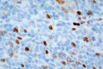
| 应用说明 | IHC-P: IHC-P was tested in the publication reference (PMID: 34650054) and sodium citrate was used as antigen retrieval buffer * The dilutions indicate recommended starting dilutions and the optimal dilutions or concentrations should be determined by the scientist. |
| 形式 | Liquid |
|---|---|
| 缓冲液 | Serum and 5 mM Sodium azide. |
| 抗菌剂 | 5 mM Sodium azide |
| 存放说明 | For continuous use, store undiluted antibody at 2-8°C for up to a week. For long-term storage, aliquot and store at -20°C or below. Storage in frost free freezers is not recommended. Avoid repeated freeze/thaw cycles. Suggest spin the vial prior to opening. The antibody solution should be gently mixed before use. |
| 注意事项 | For laboratory research only, not for drug, diagnostic or other use. |
| 数据库连接 | |
|---|---|
| 基因名称 | MKI67 |
| 全名 | marker of proliferation Ki-67 |
| 背景介绍 | Ki-67 is a nuclear protein. It is associated with and may be necessary for cellular proliferation. Alternatively spliced transcript variants have been described. A related pseudogene exists on chromosome X. [provided by RefSeq, Mar 2009] |
| 生物功能 | Ki-67 required to maintain individual mitotic chromosomes dispersed in the cytoplasm following nuclear envelope disassembly (PubMed:27362226). Associates with the surface of the mitotic chromosome, the perichromosomal layer, and covers a substantial fraction of the chromosome surface (PubMed:27362226). Prevents chromosomes from collapsing into a single chromatin mass by forming a steric and electrostatic charge barrier: the protein has a high net electrical charge and acts as a surfactant, dispersing chromosomes and enabling independent chromosome motility (PubMed:27362226). Binds DNA, with a preference for supercoiled DNA and AT-rich DNA (PubMed:10878551). Does not contribute to the internal structure of mitotic chromosomes. May play a role in chromatin organization (PubMed:24867636). It is however unclear whether it plays a direct role in chromatin organization or whether it is an indirect consequence of its function in maintaining mitotic chromosomes dispersed (Probable). [UniProt] |
| 细胞定位 | Chromosome. Nucleus, nucleolus. Note=Associates with the surface of the mitotic chromosome, the perichromosomal layer, and covers a substantial fraction of the mitotic chromosome surface. Associates with satellite DNA in G1 phase. Binds tightly to chromatin in interphase, chromatin-binding decreases in mitosis when it associates with the surface of the condensed chromosomes. [UniProt] |
| 研究领域 | Microvascular Density Study antibody; Proliferation Marker antibody |
| 预测分子量 | 359 kDa |
| 翻译后修饰 | Phosphorylated. Hyperphosphorylated in mitosis. Hyperphosphorylated form does not bind DNA. [UniProt] |
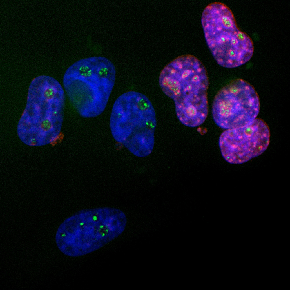
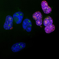
ARG11083 anti-Ki-67 antibody ICC/IF image
Immunofluorescence: HeLa cells stained with ARG11083 anti-Ki-67 antibody (red) at 1:5000 dilution and a mouse monoclonal antibody to fibrillarin at 1:2000 dilution (green). DAPI (blue) for nuclear staining.
The Ki67 protein accumulates in and around the nucleoli of interphase cells such as those on the right, and the nucleoli are revealed by the fibrillarin antibody. In contrast, cells in the quiescent G0 state such as those on the left are Ki67 negative but fibrillarin positive.
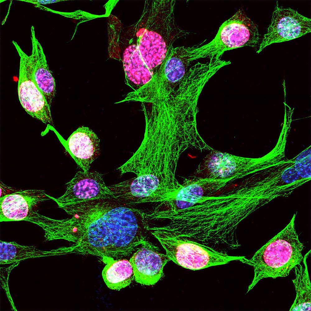
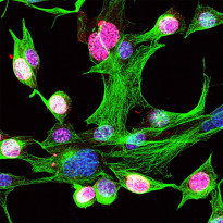
ARG11083 anti-Ki-67 antibody ICC/IF image
Immunofluorescence: NIH/3T3 cells stained with ARG11083 anti-Ki-67 antibody (red) at 1:5000 dilution and Mouse mAb to beta Tubulin (green) at 1:2000 dilution. Hoechst (blue) for nuclear staining.
The Ki-67 antibody strongly stains the nuclei of dividing cells, but not quiescent cells.
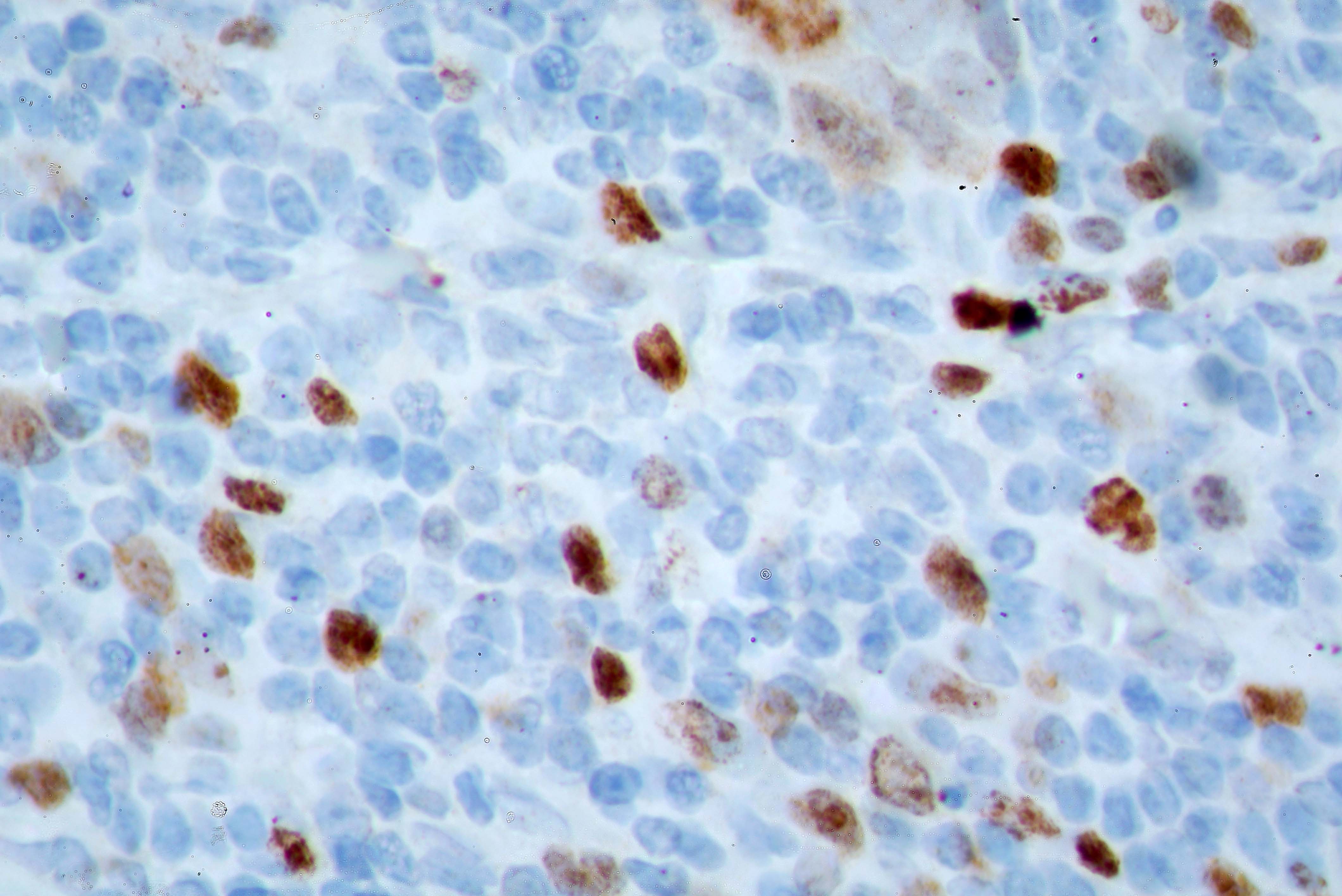
ARG11083 anti-Ki-67 antibody IHC-Fr image
Immunohistochemistry: Frozen section of Human breast tissue including both normal and cancer cells stained with ARG11083 anti-Ki-67 antibody.
The cancer cells divide rapidly and heavily express Ki-67 and so stain strongly with the Ki-67 antibody.
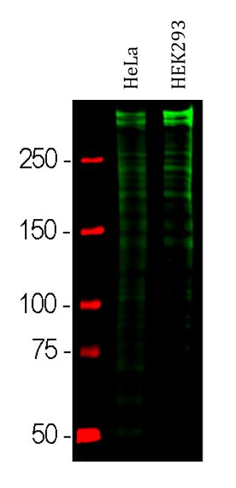
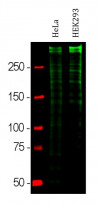
ARG11083 anti-Ki-67 antibody WB image
Western blot: HeLa and HEK293 cell lysates stained with ARG11083 anti-Ki-67 antibody at 1:10000 dilution.
Strong double bands larger than the 250 kDa standard correspond to full length 345 kDa and 395 kDa Ki67 isoforms, while smaller proteolytic fragments of these isoforms are also invariably detected on the blot.
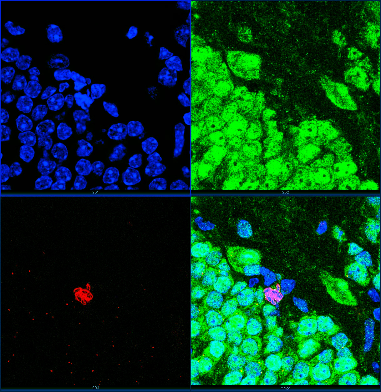
ARG11083 anti-Ki-67 antibody IHC-Fr image
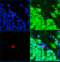
Immunohistochemistry: Frozen section of adult mouse hippocampus dentate region stained with ARG11083 anti-Ki-67 antibody (red) at 1:2000 dilution and mouse monoclonal antibody to FOX3 / NeuN (green) at 1:2000 dilution. Hoechst dye (blue) staining of DNA. Bottom right image: All three merged.
Dividing cells are very rare in adult animals, but one can be seen in the center of the image. Chromosomes can be seen in blue and their Ki-67 coating can be seen in red. The dividing cell is FOX3 / NeuN negative and so is presumably a glial cell.
 New Products


