Loading Controls for Cytoplasmic / Nuclear Fractions Antibody Panel
| 货号 | 内含物名称 | 宿主克隆性 | 反应 | 应用 | 包装 |
|---|---|---|---|---|---|
| ARG54003 | anti-COX4 antibody | Mouse mAb | Hu, Ms, Rat, Goat, Hm, Mk | FACS, ICC/IF, IHC-P, IP, WB | 25 μl |
| ARG65681 | anti-Histone H3 antibody | Mouse mAb | Hu, Ms, Rat | ICC/IF, IHC-P, IP, WB | 25 μg |
| ARG10112 | anti-GAPDH antibody [6C5] | Mouse mAb | Hu, Ms, Rat, AGMK, Bb, Cat, Chk, Dog, Fsh, Hm, Mk, Pig, Rb, Xenopus laevis, Zfsh | ELISA, ICC/IF, IHC-Fr, WB | 25 μg |
| ARG65350 | Goat anti-Mouse IgG antibody (HRP) | Goat pAb | Ms | ELISA, IHC-P, WB | 50 μl |
| 靶点名称 | Loading Controls for Cytoplasmic / Nuclear Fractions |
|---|---|
| 別名 | Loading Controls for Cytoplasmic / Nuclear Fractions antibody; GAPDH antibody; COX4 antibody; Histone H3 antibody |
| 存放说明 | For continuous use, store undiluted antibody at 2-8°C for up to a week. For long-term storage, aliquot and store at -20°C or below. Storage in frost free freezers is not recommended. Avoid repeated freeze/thaw cycles. Suggest spin the vial prior to opening. The antibody solution should be gently mixed before use. |
|---|---|
| 注意事项 | For laboratory research only, not for drug, diagnostic or other use. |
| 全名 | Antibody Panel for Loading Controls for Cytoplasmic / Nuclear Fractions |
|---|---|
| 研究领域 | Cancer antibody; Controls and Markers antibody; Gene Regulation antibody; Immune System antibody; Metabolism antibody; Neuroscience antibody; Signaling Transduction antibody |
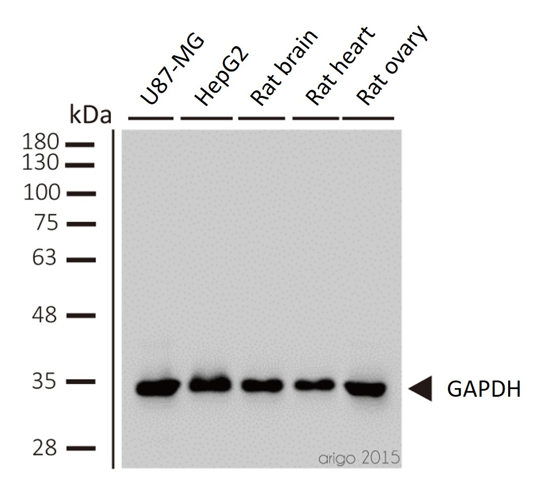
ARG10112 anti-GAPDH antibody [6C5] WB image
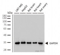
Western blot: 1) U87-MG 2) HepG2 3) rat brain 4) rat heart 5) rat ovary stained with ARG10112 anti-GAPDH antibody [6C5] at 1:2000 dilution.
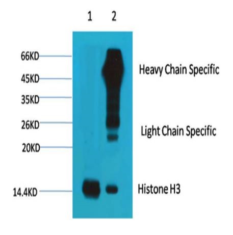
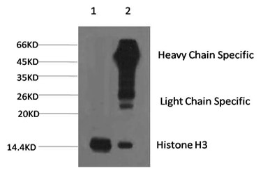
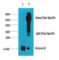
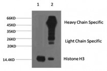
ARG65681 anti-Histone H3 antibody IP image
Immunoprecipitation: 1) HeLa cell lysate stained with ARG65681 anti-Histone H3 antibody and 2) IP product immunoprecipitated by ARG65681 anti-Histone H3 antibody at 1:200 dilution.
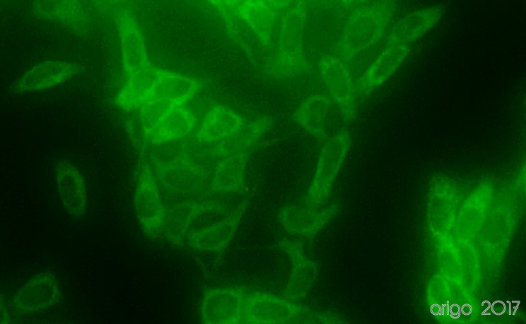
ARG10112 anti-GAPDH antibody [6C5] ICC/IF image
Immunofluorescence: 100% Methanol fixed (RT, 10 min) HeLa cells stained with ARG10112 anti-GAPDH antibody [6C5] (green) at 1:200 dilution.
Secondary antibody: ARG55393 Goat anti-Mouse IgG (H+L) antibody (FITC)
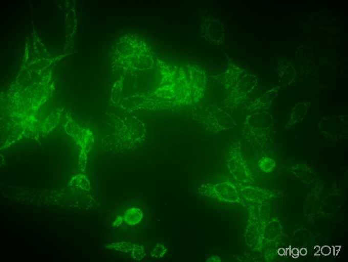
ARG54003 anti-COX4 antibody ICC/IF image
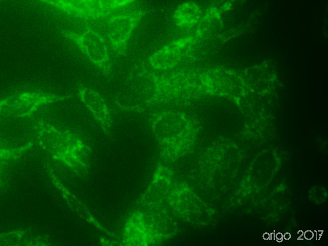
Immunofluorescence: 100% Methanol fixed (RT, 10 min) HeLa cells stained with ARG54003 anti-COX4 antibody (green) at 1:150 dilution.
Secondary antibody: ARG55393 Goat anti-Mouse IgG (H+L) antibody (FITC)
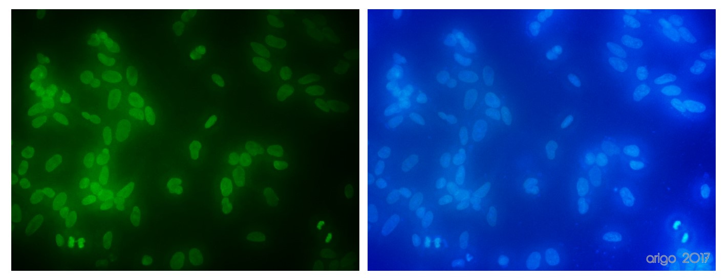
ARG65681 anti-Histone H3 antibody ICC/IF image
Immunofluorescence: 100% Methanol fixed (RT, 10 min) HeLa cells stained with ARG65681 anti-Histone H3 antibody at 1:100 dilution. Left: primary antibody (green). Right: Merge (primary antibody and DAPI).
Secondary antibody: ARG55393 Goat anti-Mouse IgG (H+L) antibody (FITC)
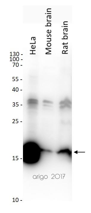
ARG65681 anti-Histone H3 antibody WB image
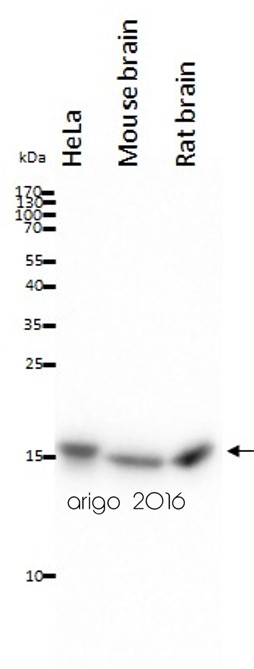
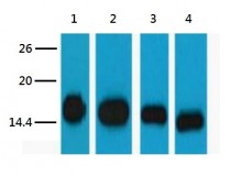
Western blot: 20 µg of HeLa, Mouse brain and Rat brain lysates stained with ARG65681 anti-Histone H3 antibody at 1:2000 dilution.
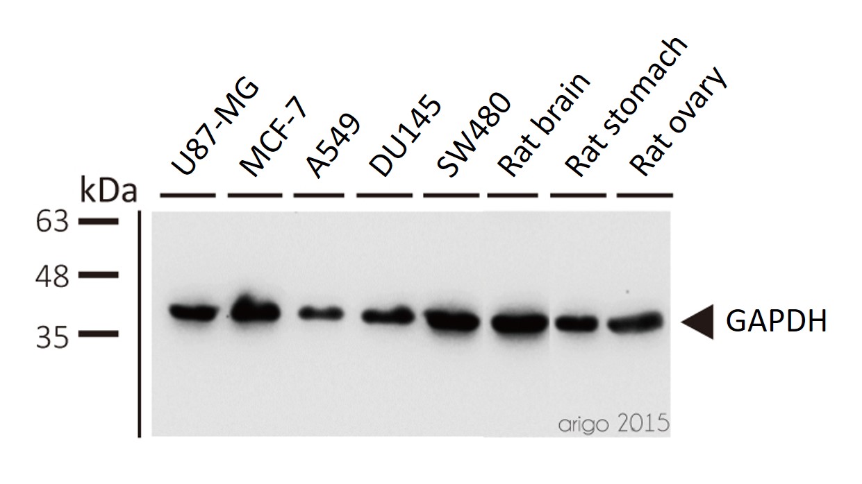
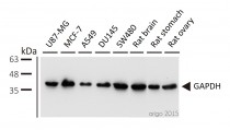
ARG10112 anti-GAPDH antibody [6C5] WB image
Western blot: 1) U87-MG 2) MCF-7 3) A549 4) DU145 5) SW480 6) rat brain 7) rat stomach 8) rat ovary stained with ARG10112 anti-GAPDH antibody [6C5] at 1:5000 dilution.
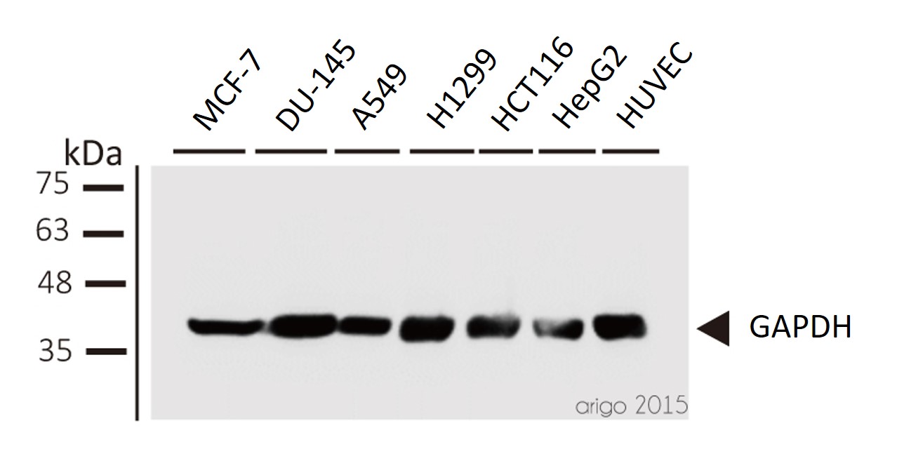
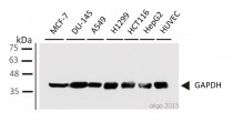
ARG10112 anti-GAPDH antibody [6C5] WB image
Western blot: 1) MCF-7 2) DU-145 3) A549 4) H1299 5) HCT116 6) HepG2 7) HUVEC stained with ARG10112 anti-GAPDH antibody [6C5] at 1:1000 dilution.
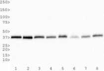
ARG10112 anti-GAPDH antibody [6C5] WB image
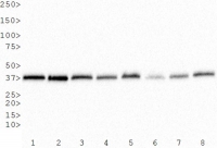
Western Blot: 1) HeLa, 2) NTERA-2, 3) A-431, 4) HepG2, 5) MCF-7, 6) NIH 3T3, 7) PC-12 and 8) COS-7 whole cell lysates stained with anti-GAPDH antibody [6C5] (ARG10112)
ARG65681 anti-Histone H3 antibody WB image
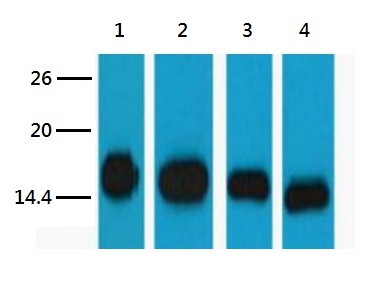
Western blot: 1) HeLa, 2) Raw, 3) Mouse brain tissue, and 4) Rat brain tissue lysates stained with ARG65681 anti-Histone H3 antibody at 1:5000 dilution.
ARG54003 anti-COX4 antibody FACS image
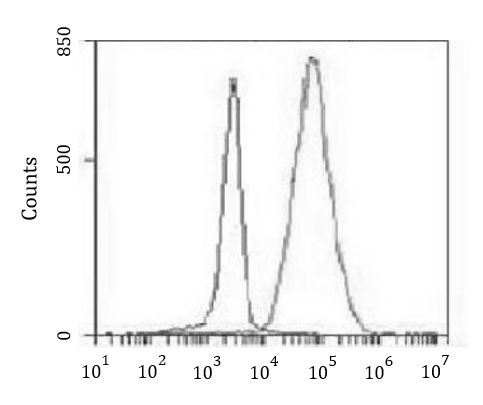
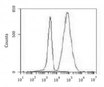
Flow Cytometry: K562 cells stained with ARG54003 anti-COX4 antibody at 1:100 dilution (right histogram) or isotype control (left histogram), followed by incubation with FITC labelled secondary antibody.
ARG54003 anti-COX4 antibody IHC-P image
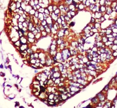
Immunohistochemistry: Paraffin-embedded Human colorectal carcinoma stained with ARG54003 anti-COX4 antibody at 1:50 dilution. Antigen Retrieval: High-pressure and temperature Citrate buffer (pH 6.0).
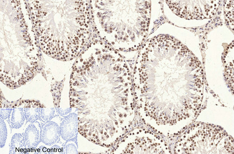
ARG65681 anti-Histone H3 antibody IHC-P image
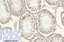
Immunohistochemistry: Paraffin-embedded Rat testis tissue stained with ARG65681 anti-Histone H3 antibody at 1:200 dilution (4°C, overnight). Antigen Retrieval: Boil tissue section in Sodium citrate buffer (pH 6.0) for 20 min. Secondary antibody was diluted at 1:200 (RT, 30 min).
Negative control was used by secondary antibody only.
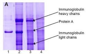
ARG10112 anti-GAPDH antibody [6C5] IP image
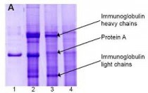
Immunoprecipitation and western blot: 1) GAPDH (1 μg). 2) GAPDH IP from rat heart tissue extract. 3) Only GAPDH preincubated with Protein A Sepharose. 4) Only Protein A Sepharose stained with ARG10112 GAPDH antibody [6C5].
Mixture of protein A-Sepharose with ARG10112 anti-GAPDH and tissue extract was incubated for 30 min at room temperature and precipitated by centrifugation. Pellet was washed with PBS, suspended in reducing electrophoresis sample buffer and heated for 5 minutes at 100ºC. After centrifugation supernatant was loaded on gel and proteins were separated by SDS electrophoresis.ARG65681 anti-Histone H3 antibody IP image
Immunoprecipitation: 1) HeLa cell lysate stained with ARG65681 anti-Histone H3 antibody and 2) IP product immunoprecipitated by ARG65681 anti-Histone H3 antibody at 1:200 dilution.
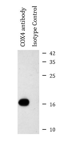
ARG54003 anti-COX4 antibody IP image
Immunoprecipitation: HeLa cell lysates were immunoprecipitated and stained with ARG54003 anti-COX4 antibody.
ARG54003 anti-COX4 antibody WB image
Western blot: 20 µg of HeLa, Mouse brain and Rat brain lysates stained with ARG54003 anti-COX4 antibody at 1:1000 dilution.
ARG54003 anti-COX4 antibody ICC/IF image
Immunofluorescence: 100% Methanol fixed (RT, 10 min) HeLa cells stained with ARG54003 anti-COX4 antibody (green) at 1:150 dilution.
Secondary antibody: ARG55393 Goat anti-Mouse IgG (H+L) antibody (FITC)
ARG10112 anti-GAPDH antibody [6C5] WB image
Western blot: Mouse samples stained with ARG10112 anti-GAPDH antibody [6C5] at 1:1000 dilution.
From Yun-Yun Li et al. Int J Biol Sci (2022), doi: 10.7150/ijbs.68224, Fig. 5. C.
ARG10112 anti-GAPDH antibody [6C5] WB image
Western blot: Porcine kidney stained with ARG10112 anti-GAPDH antibody [6C5].
From Jianni Huang et al. Front Cell Dev Biol (2022), doi: 10.3389/fcell.2022.899869, Fig. 2. E.
ARG10112 anti-GAPDH antibody [6C5] WB image
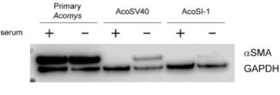
Western blot: pAFs, AcoSV40, and AcoSI-1 stained with ARG10112 anti-GAPDH antibody [6C5] at 1:5000 dilution.
From Michele N Dill et al. PLoS One. (2023), doi: 10.3389/fcell.2022.899869, Fig. 2. C.
 New Products


