NFkB nuclear translocation Antibody Panel
| 货号 | 内含物名称 | 宿主克隆性 | 反应 | 应用 | 包装 |
|---|---|---|---|---|---|
| ARG10112 | anti-GAPDH antibody [6C5] | Mouse mAb | Hu, Ms, Rat, AGMK, Bb, Cat, Chk, Dog, Fsh, Hm, Mk, Pig, Rb, Xenopus laevis, Zfsh | ELISA, ICC/IF, IHC-Fr, WB | 50 μg |
| ARG54747 | anti-Histone H3 antibody | Mouse mAb | Hu | WB | 100 μl |
| ARG54746 | anti-NFkB p105 / p50 antibody | Mouse mAb | Hu | FACS, IHC-P, WB | 100 μl |
| ARG51013 | anti-NFkB p65 antibody | Rabbit pAb | Hu, Ms, Rat | IHC-Fr, IHC-P, WB | 50 μl |
| ARG65350 | Goat anti-Mouse IgG antibody (HRP) | Goat pAb | Ms | ELISA, IHC-P, WB | 50 μl |
| 产品描述 | Nuclear factor kappa B (NFkB) is a protein complex that controls large number of genes responsible for the regulation of apoptosis, inflammation, tumorigenesis and a variety of immune responses. NF-kB is sensitive and responsive to stimuli such as cytokines, stress, UV, low oxygen, bacterial or viral infections. Upon activation, NFkB is translocated into nucleus and exert its function on the transcriptional regulation of NFkB-responsive genes. Histone H3 is a marker for nuclear fraction while GAPDH is a marker for cytosolic fraction. Baeuerle et al. 1991. Biochem Biophys Acta 1072: 63-80 Lenardo and Baltimore. 1989. Cell 58: 227 |
|---|---|
| 靶点名称 | NFkB nuclear translocation |
| 別名 | NFkB nuclear translocation antibody; GAPDH antibody; NFkB p65 antibody; NFkB p105 / p50 antibody; Histone H3 antibody |
| 存放说明 | For continuous use, store undiluted antibody at 2-8°C for up to a week. For long-term storage, aliquot and store at -20°C or below. Storage in frost free freezers is not recommended. Avoid repeated freeze/thaw cycles. Suggest spin the vial prior to opening. The antibody solution should be gently mixed before use. |
|---|---|
| 注意事项 | For laboratory research only, not for drug, diagnostic or other use. |
| 全名 | Antibody Panel for NFkB nuclear translocation |
|---|---|
| 研究领域 | Cancer antibody; Cell Biology and Cellular Response antibody; Cell Death antibody; Controls and Markers antibody; Gene Regulation antibody; Immune System antibody; Metabolism antibody; Microbiology and Infectious Disease antibody; Neuroscience antibody; Signaling Transduction antibody |
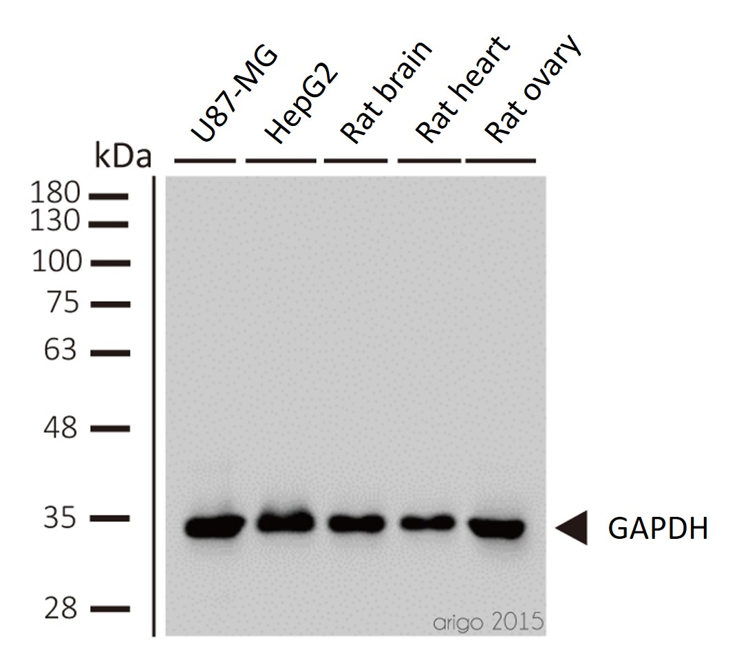
ARG10112 anti-GAPDH antibody [6C5] WB image
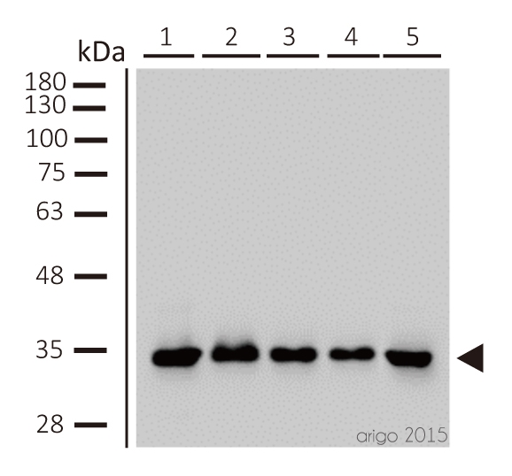
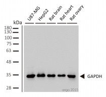
Western blot: 1) U87-MG 2) HepG2 3) rat brain 4) rat heart 5) rat ovary stained with ARG10112 anti-GAPDH antibody [6C5] at 1:2000 dilution.
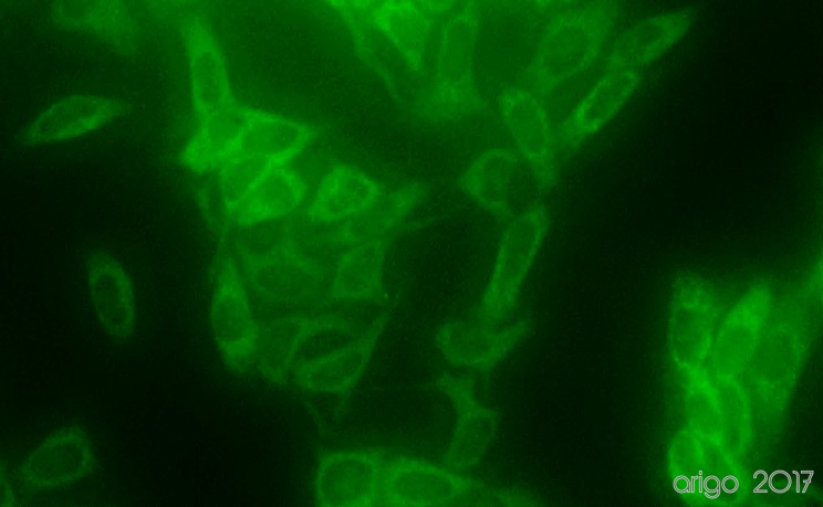
ARG10112 anti-GAPDH antibody [6C5] ICC/IF image
Immunofluorescence: 100% Methanol fixed (RT, 10 min) HeLa cells stained with ARG10112 anti-GAPDH antibody [6C5] (green) at 1:200 dilution.
Secondary antibody: ARG55393 Goat anti-Mouse IgG (H+L) antibody (FITC)
ARG54747 anti-Histone H3 antibody WB image
Western blot: 20 µg of 293T cell lysate stained with ARG54747 anti-Histone H3 antibody at 1:2000 dilution.
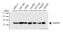
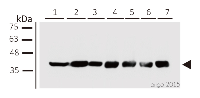
ARG10112 anti-GAPDH antibody [6C5] WB image
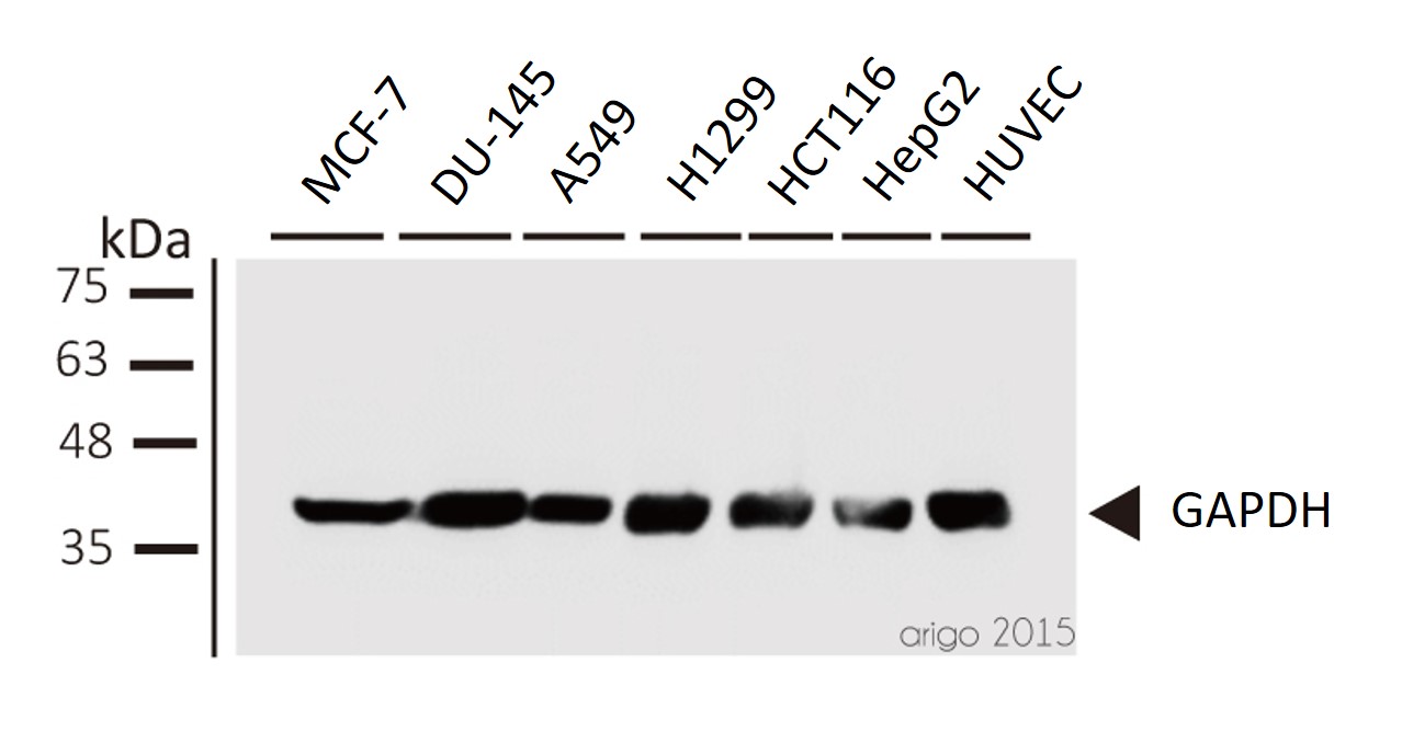
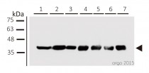
Western blot: 1) MCF-7 2) DU-145 3) A549 4) H1299 5) HCT116 6) HepG2 7) HUVEC stained with ARG10112 anti-GAPDH antibody [6C5] at 1:1000 dilution.
ARG10112 anti-GAPDH antibody [6C5] WB image
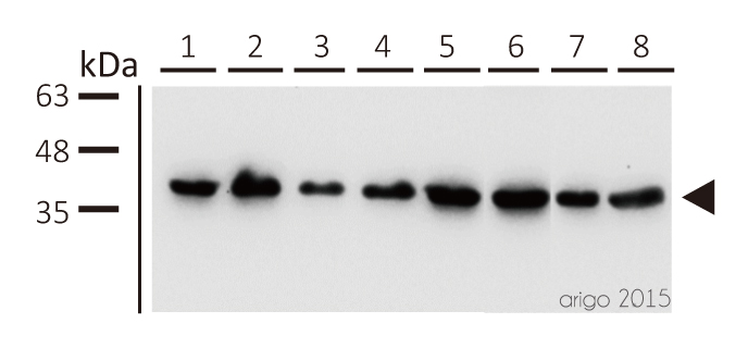
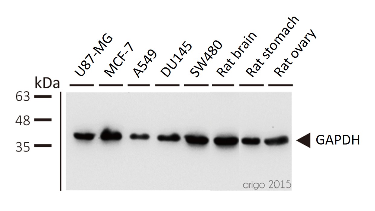
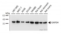
Western blot: 1) U87-MG 2) MCF-7 3) A549 4) DU145 5) SW480 6) rat brain 7) rat stomach 8) rat ovary stained with ARG10112 anti-GAPDH antibody [6C5] at 1:5000 dilution.
ARG10112 anti-GAPDH antibody [6C5] WB image
Western blot: 1) U87-MG 2) HepG2 3) rat brain 4) rat heart 5) rat ovary stained with ARG10112 anti-GAPDH antibody [6C5] at 1:2000 dilution.
ARG10112 anti-GAPDH antibody [6C5] WB image
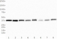
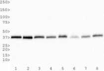
Western Blot: 1) HeLa, 2) NTERA-2, 3) A-431, 4) HepG2, 5) MCF-7, 6) NIH 3T3, 7) PC-12 and 8) COS-7 whole cell lysates stained with anti-GAPDH antibody [6C5] (ARG10112)
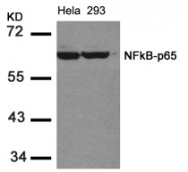
ARG51013 anti-NFkB p65 antibody WB image
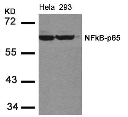
Western Blot: extracts from HeLa and 293 cells stained with anti-NFkB p65 antibody ARG51013.
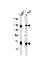
ARG54746 anti-NFkB p105 / p50 antibody WB image
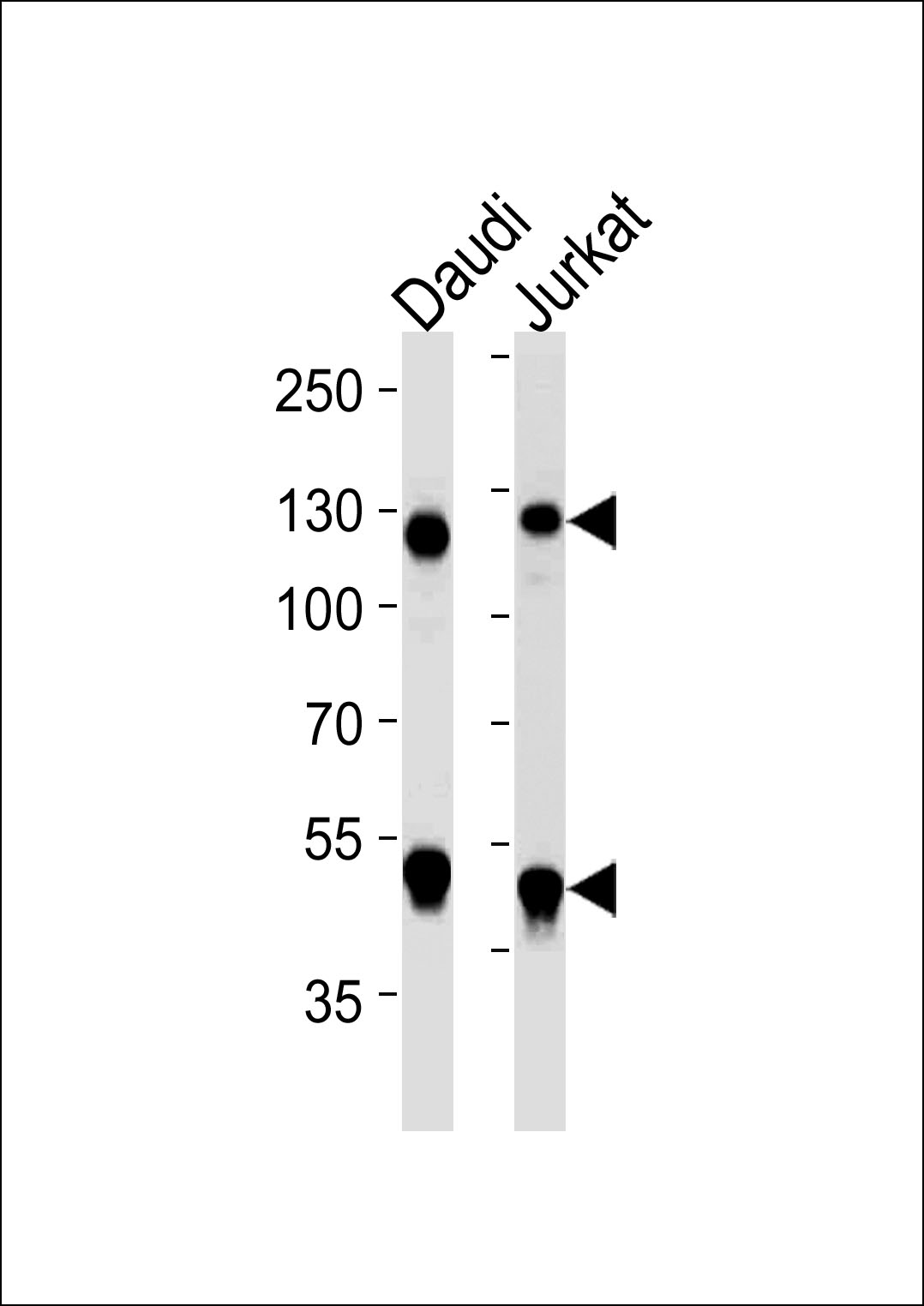
Western blot: 35 μg of Daudi, Jurkat cell line lysate (from left to right) stained with ARG54746 anti-NFkB p105 / p50 antibody at 1:1000 dilution.
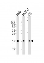
ARG54747 anti-Histone H3 antibody WB image
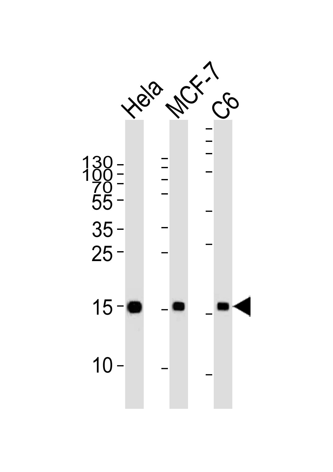
Western blot: 35 μg of HeLa, MCF-7, C6 cell line lysates (from left to right) stained with ARG54747 anti-Histone H3 antibody at 1:2000 dilution.
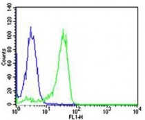
ARG54746 anti-NFkB p105 / p50 antibody FACS image
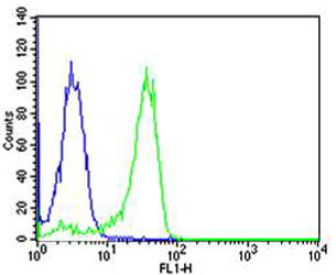
Flow Cytometry: HeLa cells stained with ARG54746 anti-NFkB p105 / p50 antibody (green) at 1:25 dilution or isotype control antibody (blue), followed by incubation with Alexa Fluor® 488 labelled secondary antibody.
ARG54746 anti-NFkB p105 / p50 antibody IHC-P image
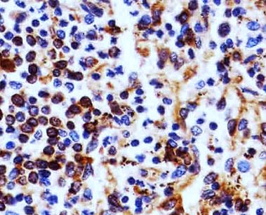
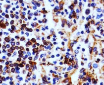
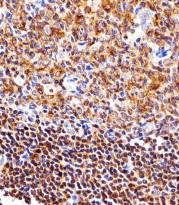
Immunohistochemistry: Paraffin-embedded Human spleen section stained with ARG54746 anti-NFkB p105 / p50 antibody at 1:25 dilution.
ARG10112 anti-GAPDH antibody [6C5] IP image
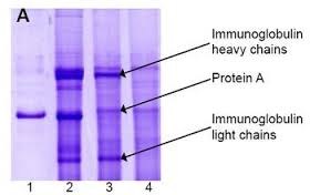
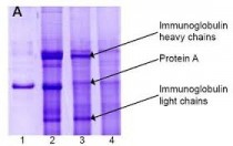
Immunoprecipitation and western blot: 1) GAPDH (1 μg). 2) GAPDH IP from rat heart tissue extract. 3) Only GAPDH preincubated with Protein A Sepharose. 4) Only Protein A Sepharose stained with ARG10112 GAPDH antibody [6C5].
Mixture of protein A-Sepharose with ARG10112 anti-GAPDH and tissue extract was incubated for 30 min at room temperature and precipitated by centrifugation. Pellet was washed with PBS, suspended in reducing electrophoresis sample buffer and heated for 5 minutes at 100ºC. After centrifugation supernatant was loaded on gel and proteins were separated by SDS electrophoresis.ARG54746 anti-NFkB p105 / p50 antibody WB image
Western blot: 35 µg of Daudi cell lysate stained with ARG54746 anti-NFkB p105 / p50 antibody at 1:1000 dilution.
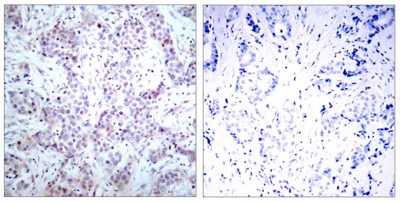
ARG51013 anti-NFkB p65 antibody IHC-P image
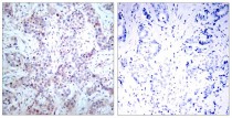
Immunohistochemistry: paraffin-embedded human breast carcinoma tissue stained with anti-NFkB p65 antibody ARG51013 (left) or the same antibody preincubated with blocking peptide (right).
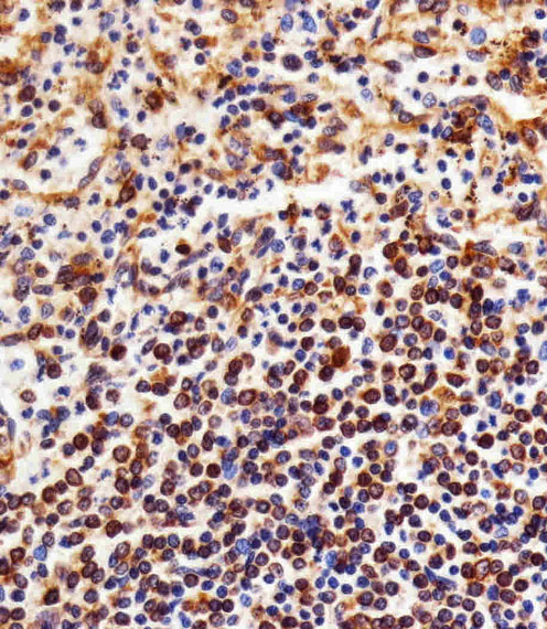
ARG54746 anti-NFkB p105 / p50 antibody IHC image
Immunohistochemistry: paraffin-embedded Human spleen section stained with ARG54746 anti-NFkB p105 / p50 antibody at 1:25 dilution.
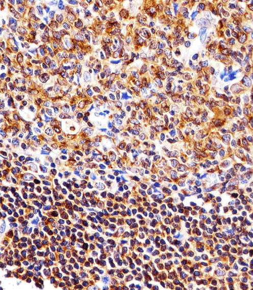
ARG54746 anti-NFkB p105 / p50 antibody IHC image
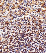
Immunohistochemistry: paraffin-embedded Human tonsil section stained with ARG54746 anti-NFkB p105 / p50 antibody at 1:25 dilution.
ARG54746 anti-NFkB p105 / p50 antibody FACS image
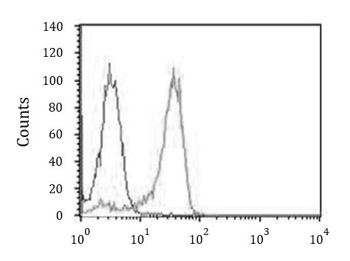
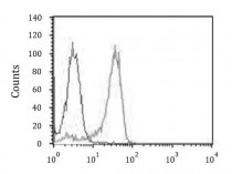
Flow Cytometry: HeLa cells stained with ARG54746 anti-NFkB p105 / p50 antibody (right histogram) at 1:25 dilution or isotype control antibody (left histogram), followed by incubation with Alexa Fluor® 488 labelled secondary antibody.
ARG10112 anti-GAPDH antibody [6C5] WB image
Western blot: 1) U87-MG 2) MCF-7 3) A549 4) DU145 5) SW480 6) rat brain 7) rat stomach 8) rat ovary stained with ARG10112 anti-GAPDH antibody [6C5] at 1:5000 dilution.
ARG51013 anti-NFkB p65 antibody IHC-Fr image
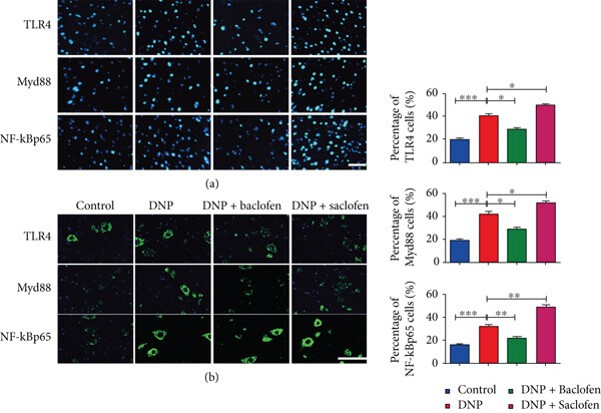
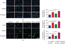
Immunofluorescence: Rat (L1–5) spinal cord stained with ARG51013 anti-NFkB p65 antibody and ARG54348 anti-MyD88 antibody.
From Peng Liu et al. Mediators of inflammation (2018), doi: .org/10.1155/2018/6016272, Fig. 5.
ARG10112 anti-GAPDH antibody [6C5] WB image
Western blot: Mouse samples stained with ARG10112 anti-GAPDH antibody [6C5] at 1:1000 dilution.
From Yun-Yun Li et al. Int J Biol Sci (2022), doi: 10.7150/ijbs.68224, Fig. 5. C.
ARG10112 anti-GAPDH antibody [6C5] WB image
Western blot: Porcine kidney stained with ARG10112 anti-GAPDH antibody [6C5].
From Jianni Huang et al. Front Cell Dev Biol (2022), doi: 10.3389/fcell.2022.899869, Fig. 2. E.
ARG10112 anti-GAPDH antibody [6C5] WB image
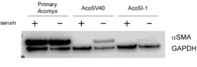
Western blot: pAFs, AcoSV40, and AcoSI-1 stained with ARG10112 anti-GAPDH antibody [6C5] at 1:5000 dilution.
From Michele N Dill et al. PLoS One. (2023), doi: 10.3389/fcell.2022.899869, Fig. 2. C.
ARG10112 anti-GAPDH antibody [6C5] WB image
Western blot: 1) MCF-7 2) DU-145 3) A549 4) H1299 5) HCT116 6) HepG2 7) HUVEC stained with ARG10112 anti-GAPDH antibody [6C5] at 1:1000 dilution.
 New Products


