Tag Internal Control Antibody Duo (GFP, GAPDH)
| 货号 | 内含物名称 | 宿主克隆性 | 反应 | 应用 | 包装 |
|---|---|---|---|---|---|
| ARG10112 | anti-GAPDH antibody [6C5] | Mouse mAb | Hu, Ms, Rat, AGMK, Bb, Cat, Chk, Dog, Fsh, Hm, Mk, Pig, Rb, Xenopus laevis, Zfsh | ELISA, ICC/IF, IHC-Fr, WB | 50 μg |
| ARG62343 | anti-GFP antibody [GF28R] | Mouse mAb | Other | Dot, ELISA, ICC/IF, IP, WB | 50 μg |
| 产品描述 | GFP has become one of the most important tools used in bioscience. When using GFP as a reporter for protein expressing study, arigo recommend to include GAPDH as a loading control to normalize the sample loading and the experimental errors to show the convincing result. GAPDH is often stably and constitutively expressed at high levels in most tissues and cells in cytoplasm. Therefore, GAPDH is a widely used loading control and cytoplasm marker for western blot (35-40kDa) and ICC/IF study. Usage note: S-nitrosylated GAPDH might translocate to nucleus especially for nitric oxide-related studies. In addition, the GAPDH expression level is up regulated under hypoxia condition. Therefore, GAPDH is not suitable for loading control and cytoplasm marker for oxygen-related and nitric oxide-related studies. GFP, green fluorescent protein, is a protein from jellyfish Aequorea Victoria with 238 amino acids residues (26.9 kDa) that exhibits bright green fluorescence when exposed to light in the blue to ultraviolet range. Arigo ARG62344 GFP antibody recognizes native and denatured forms of GFP and its variants such as: EGFP, YFP, EYFP, and CFP. |
|---|---|
| 靶点名称 | Tag Internal Control |
| 別名 | Tag Internal Control antibody; GAPDH antibody; GFP antibody |
| 存放说明 | For continuous use, store undiluted antibody at 2-8°C for up to a week. For long-term storage, aliquot and store at -20°C or below. Storage in frost free freezers is not recommended. Avoid repeated freeze/thaw cycles. Suggest spin the vial prior to opening. The antibody solution should be gently mixed before use. |
|---|---|
| 注意事项 | For laboratory research only, not for drug, diagnostic or other use. |
| 全名 | Antibody Duo for Tag Internal Control (GFP, GAPDH) |
|---|---|
| 研究领域 | Cancer antibody; Controls and Markers antibody; Gene Regulation antibody; Metabolism antibody; Neuroscience antibody; Signaling Transduction antibody |
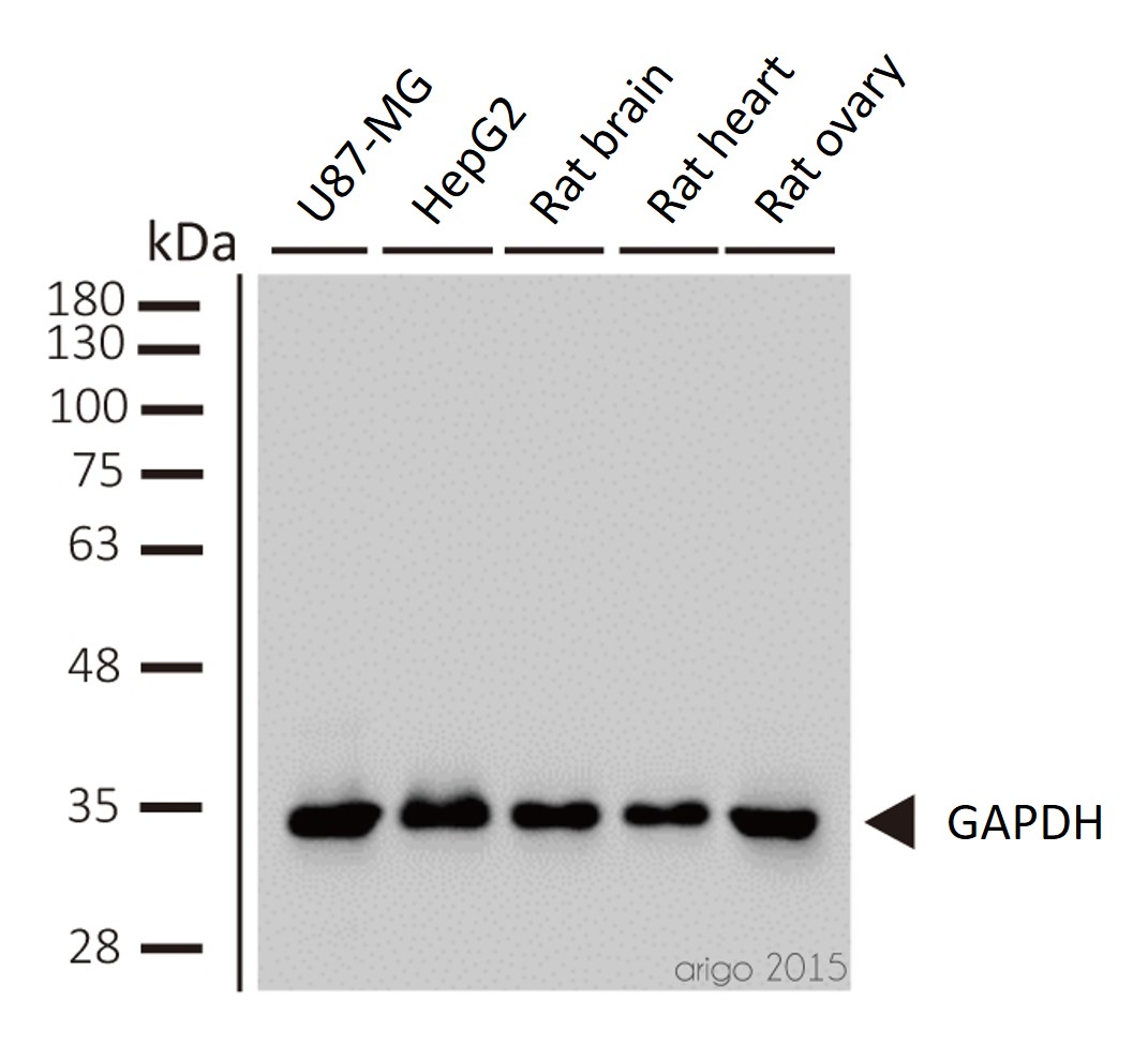
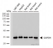
ARG10112 anti-GAPDH antibody [6C5] WB image
Western blot: 1) U87-MG 2) HepG2 3) rat brain 4) rat heart 5) rat ovary stained with ARG10112 anti-GAPDH antibody [6C5] at 1:2000 dilution.
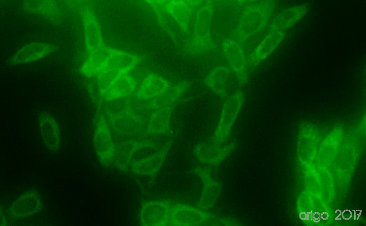
ARG10112 anti-GAPDH antibody [6C5] ICC/IF image
Immunofluorescence: 100% Methanol fixed (RT, 10 min) HeLa cells stained with ARG10112 anti-GAPDH antibody [6C5] (green) at 1:200 dilution.
Secondary antibody: ARG55393 Goat anti-Mouse IgG (H+L) antibody (FITC)
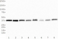
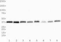
ARG10112 anti-GAPDH antibody [6C5] WB image
Western Blot: 1) HeLa, 2) NTERA-2, 3) A-431, 4) HepG2, 5) MCF-7, 6) NIH 3T3, 7) PC-12 and 8) COS-7 whole cell lysates stained with anti-GAPDH antibody [6C5] (ARG10112)
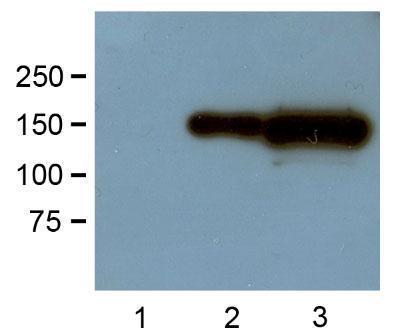
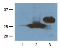
ARG62343 anti-GFP antibody [GF28R] WB image
Western Blot: HEK293 cells transfected with GFP-tagged protein vector; (1) untransfected and (2) transfected stained with ARG62343 anti-GFP antibody [GF28R] at 1:1000 (1 μg/mL) dilution
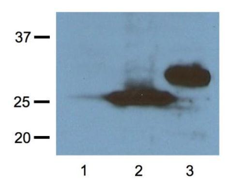
ARG62344 anti-RFP antibody [RF5R] WB image
Western Blot: HEK293 cells transfected with RFP-tagged protein vector; (1) untransfected and (2) transfected with Turbo-RFP (2), and (3) transfected with DsRed stained with ARG62344 anti-RFP antibody [RF5R] at 1:1000 (1 μg/mL) dilution
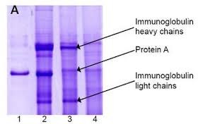
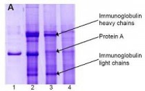
ARG10112 anti-GAPDH antibody [6C5] IP image
Immunoprecipitation and western blot: 1) GAPDH (1 μg). 2) GAPDH IP from rat heart tissue extract. 3) Only GAPDH preincubated with Protein A Sepharose. 4) Only Protein A Sepharose stained with ARG10112 GAPDH antibody [6C5].
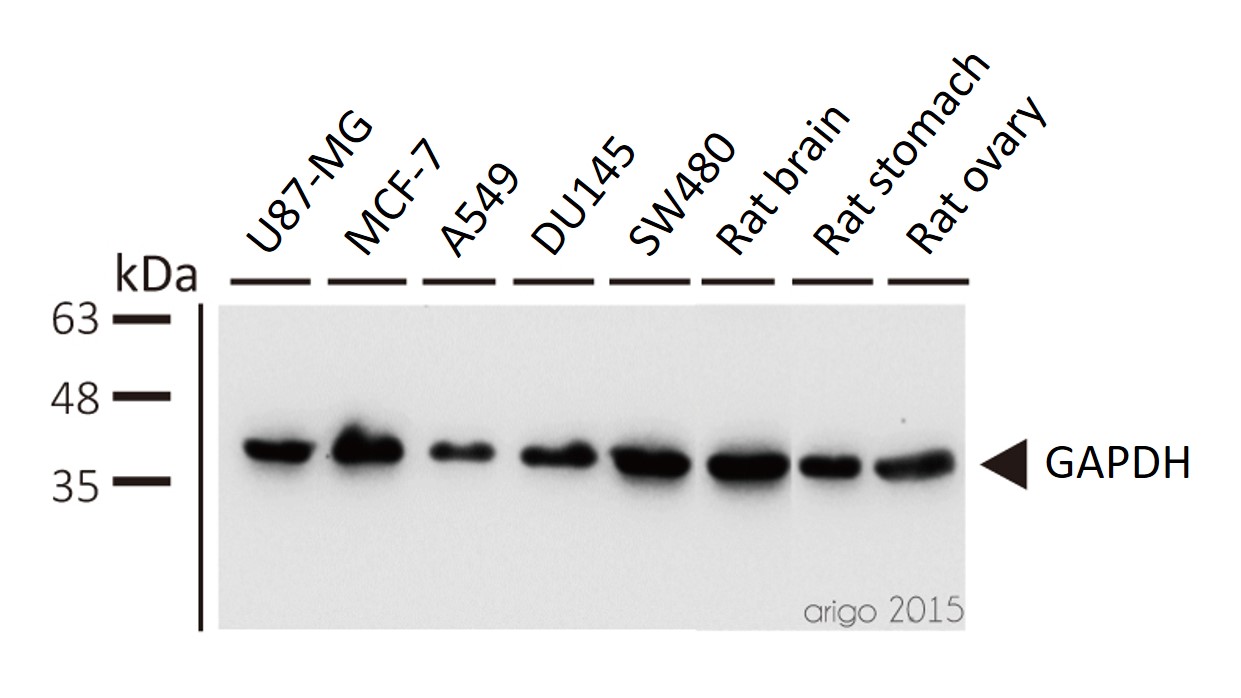
Mixture of protein A-Sepharose with ARG10112 anti-GAPDH and tissue extract was incubated for 30 min at room temperature and precipitated by centrifugation. Pellet was washed with PBS, suspended in reducing electrophoresis sample buffer and heated for 5 minutes at 100ºC. After centrifugation supernatant was loaded on gel and proteins were separated by SDS electrophoresis.ARG10112 anti-GAPDH antibody [6C5] WB image
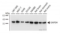
Western blot: 1) U87-MG 2) MCF-7 3) A549 4) DU145 5) SW480 6) rat brain 7) rat stomach 8) rat ovary stained with ARG10112 anti-GAPDH antibody [6C5] at 1:5000 dilution.
ARG10112 anti-GAPDH antibody [6C5] WB image
Western blot: Mouse samples stained with ARG10112 anti-GAPDH antibody [6C5] at 1:1000 dilution.
From Yun-Yun Li et al. Int J Biol Sci (2022), doi: 10.7150/ijbs.68224, Fig. 5. C.
ARG10112 anti-GAPDH antibody [6C5] WB image
Western blot: Porcine kidney stained with ARG10112 anti-GAPDH antibody [6C5].
From Jianni Huang et al. Front Cell Dev Biol (2022), doi: 10.3389/fcell.2022.899869, Fig. 2. E.
ARG10112 anti-GAPDH antibody [6C5] WB image
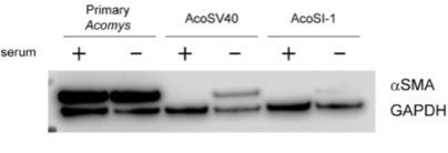
Western blot: pAFs, AcoSV40, and AcoSI-1 stained with ARG10112 anti-GAPDH antibody [6C5] at 1:5000 dilution.
From Michele N Dill et al. PLoS One. (2023), doi: 10.3389/fcell.2022.899869, Fig. 2. C.
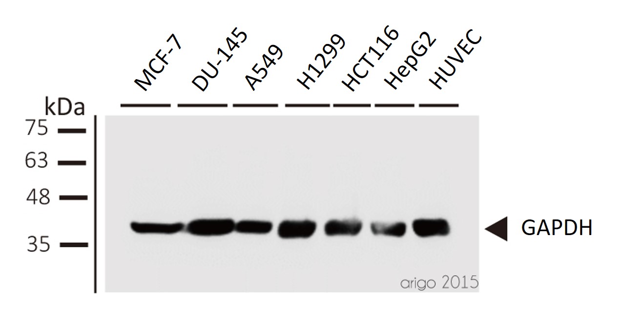
ARG10112 anti-GAPDH antibody [6C5] WB image
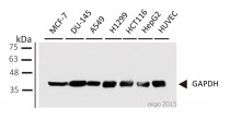
Western blot: 1) MCF-7 2) DU-145 3) A549 4) H1299 5) HCT116 6) HepG2 7) HUVEC stained with ARG10112 anti-GAPDH antibody [6C5] at 1:1000 dilution.
 New Products


